Download Coronary Sinus Real Heart
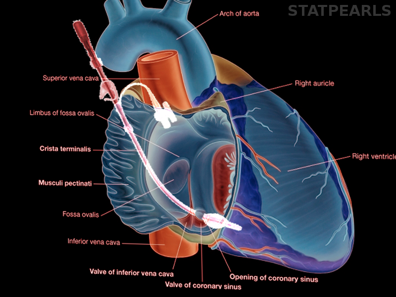
Background. In this image, you will find aorta, left pulmonary artery, left pulmonary veins, auricle of left atrium, left atrium, great cardiac vein, posterior vein of left ventricle, left ventricle, apex, superior vena cava, right pulmonary artery, right pulmonary veins, right atrium, inferior vena cava, coronary sinus. The coronary sinus is roughly a 3 cm saccular dilatation between the left cardiac chambers.
Retrograde Cardioplegia Article from www.statpearls.com
Unroofed coronary sinus (ucs) is among the rarest congenital heart malformations in pediatric practice. Coronary sinus sup vena cava inf vena cava. The coronary sinus is a collection of smaller veins that merge together to form the sinus (or large vessel), which is located along the heart's posterior (rear) surface between the left ventricle and left atrium.
Learn vocabulary, terms and more with flashcards, games and other study tools.
The coronary arteries arise from the coronary sinuses immediately distal (superior) to the aortic valve and supply the myocardium with oxygenated blood. While it's helpful to be aware. Partly overlying the cardiac crux. It is oriented obliquely in the posterior atrioventricular groove;