48+ Coronary Sinus Type Asd Photos. At the entrance of both the vcs and the vci. Individuals with a sinus venosus asd exhibit a left axis deviation of the p wave (not the qrs complex).

It is present in all mammals, including humans.
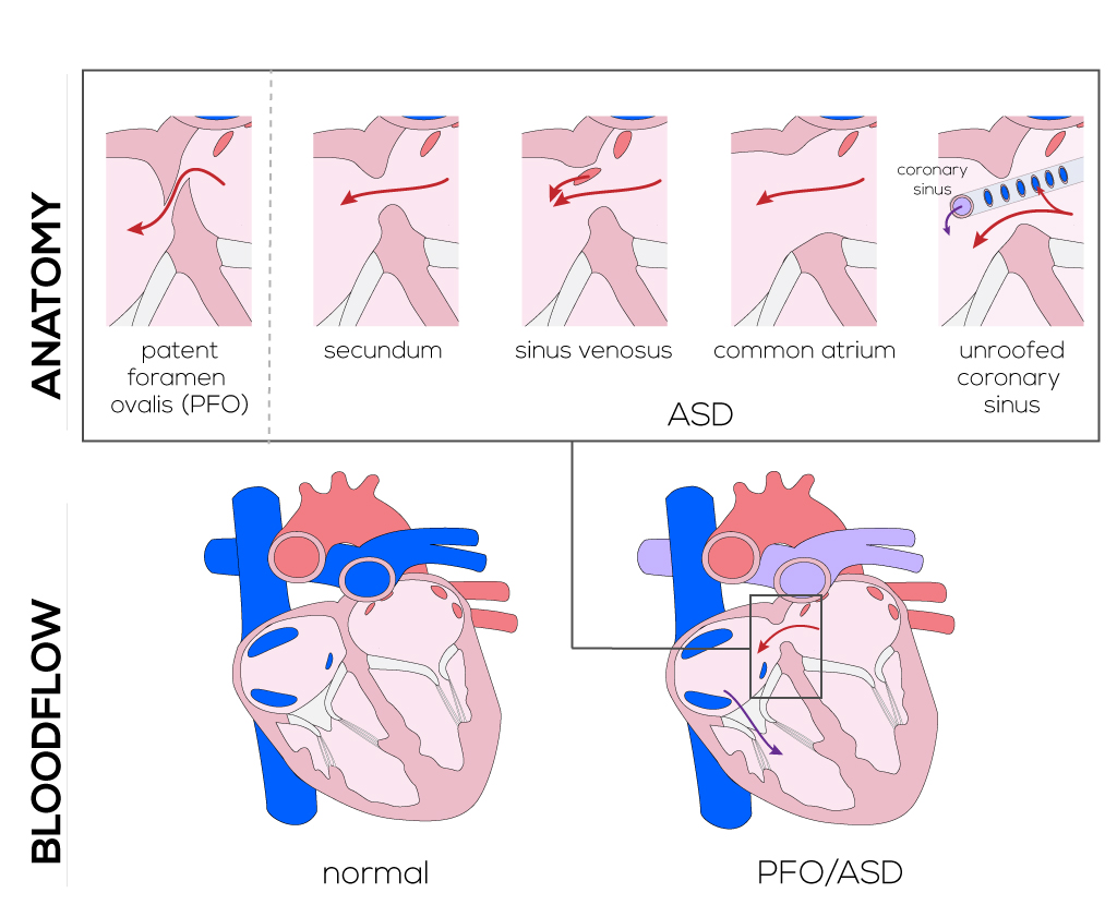
Asds are classified into septum primum, septum secundum, sinus venosus, and coronary sinus defects based on their anatomic locations. However, if the asd is large, permitting a large outcomes also depend on the type of asd, as well as how early in life the asd was diagnosed and. The coronary sinus is a collection of veins joined together to form a large vessel that collects blood from the heart muscle (myocardium). Although device erosion of the dilated coronary sinus is suspected, the defect in the coronary sinus may have been present prior to asd device closure.