32+ Femoral Head Ossification Center Appearance Images
28/11/2001 00:00
32+ Femoral Head Ossification Center Appearance
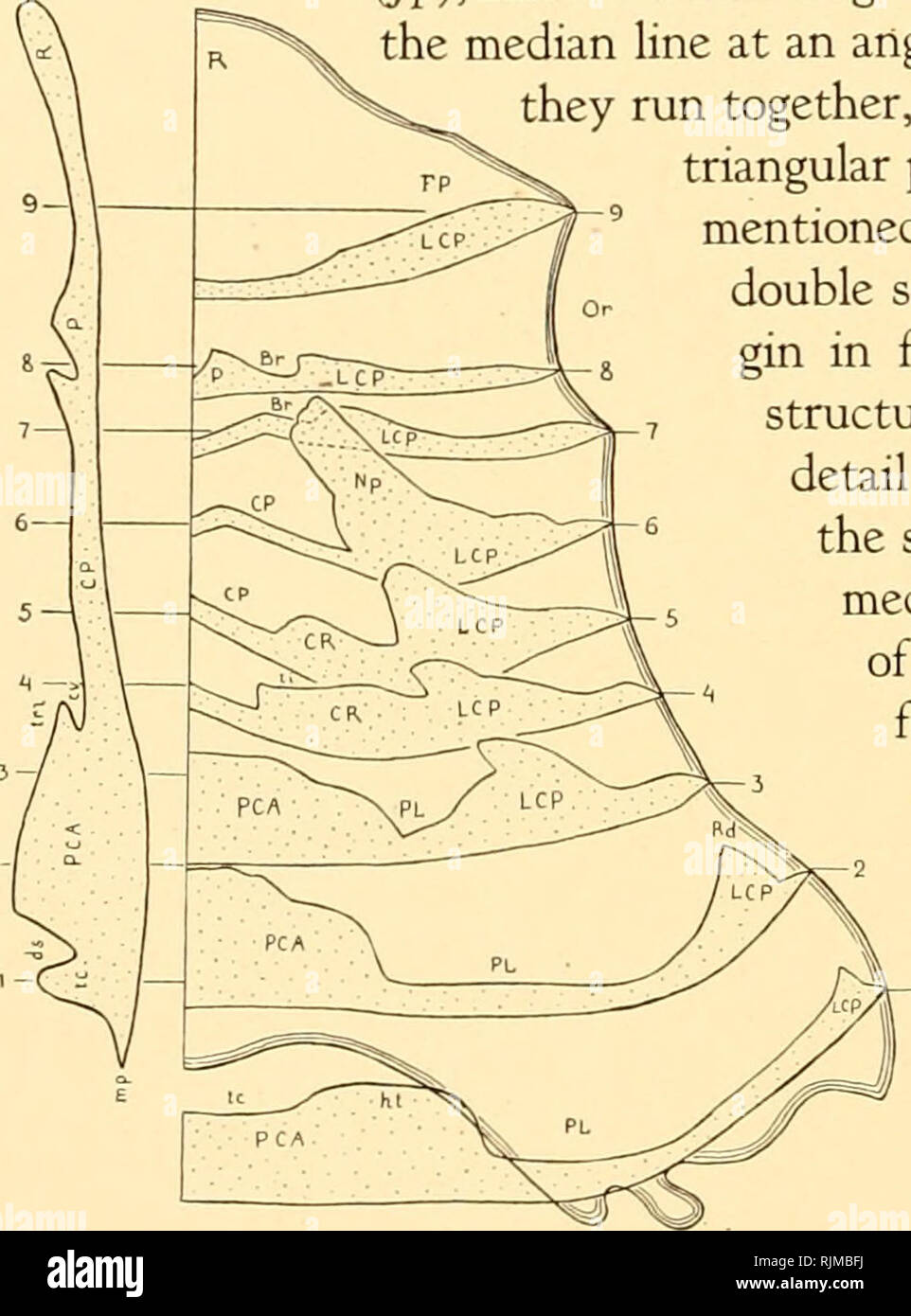
Images. Knowledge of the normal sonographic appearance of the femoral head ossification center by age and ethnicity will help clinicians in the. Ossification in the acetabular cup begins from two separate centers (os acetabuli) between the ilium and pubis, and between the ilium and ischium.
Ossification Center High Resolution Stock Photography And Images Alamy from c8.alamy.com
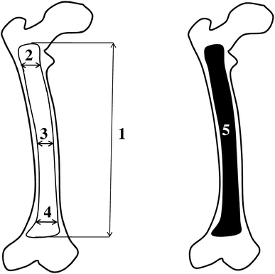
The femur ossifies from 5 centers: The femoral head is at particular risk of developing avascular necrosis because there is an area of reduced vascularization (watershed zone) between the foveolar artery is the main artery implicated in avascular necrosis of the femoral head. On ossification centers in human embryos less than one hundred days old.
The ossification center was noted at 2 weeks of life in the israeli infants and at 8 weeks in the indian infants.
The femoral head is at particular risk of developing avascular necrosis because there is an area of reduced vascularization (watershed zone) between the foveolar artery is the main artery implicated in avascular necrosis of the femoral head. Shenton lines (s) are continuous and demarcated by the dashed lines. Ossification in the acetabular cup begins from two separate centers (os acetabuli) between the ilium and pubis, and between the ilium and ischium. What is the secondary ossification center, when does it occur and where is it found?