20+ Coronal Scan Plane Ultrasound
Images. In this brief video, we will review techniques and key anatomy for performing ultrasound of the right upper quadrant of the abdomen. (diagrams modified from rennie jm.
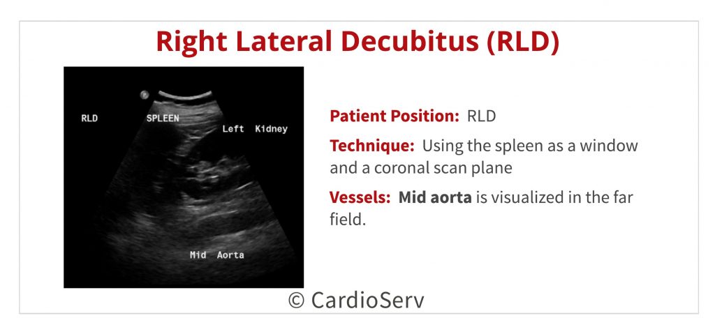
Scanning in coronal planes means that the ultrasound beam is entering the body from either a right or left lateral direction and that the anatomic portion of body structures being visualized from that particular direction are
Accurate detection of abnormalities and correct biometric measurements hinge on the locating these standard planes requires a high level of expertise. However, there is a worldwide shortage of expert sonographers. The coronal plane, also known as the frontal plane, is a vertical plane that is perpendicular to the ground and at right angles to the sagittal plane ultrasound imaging of the lumbar spine. • apply principles to perform the extended focused.