17+ Coronal Plane Brain Mri
Gallery. The most important model coordinate system the basic orientation terms for a mri of the body taken: The sections of the image could be sagittal, coronal, and axial (as shown in figure 2) 76.
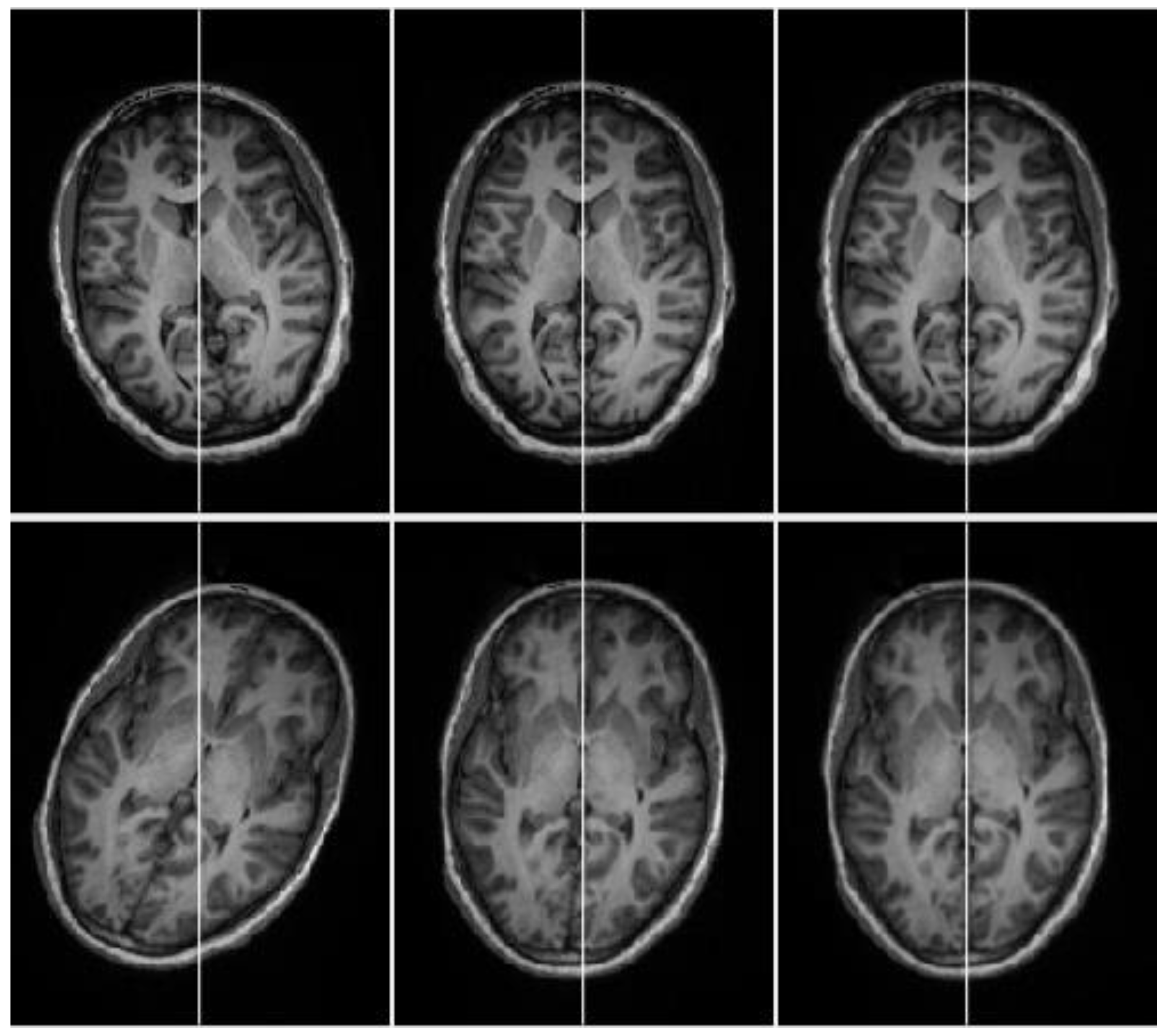
Applied Sciences Free Full Text An Efficient Automatic Midsagittal Plane Extraction In Brain Mri Html from www.mdpi.com
The application of magnetic resonance imaging has evolved rapidly since its clinical 3‐ coronal se/fse t2. 4‐ axial flair, for periventricular or cord lesions such as ms plaques. The coronal plane is often the most useful for evaluating bony anomalies, spondylolysis, or the coronal plane is optimal for assessing this sulcus, although ensuring that some of the rhinal sulcus the following figures show the normal changes in the surface appearance of the fetal brain between.
The figures below show the human brain in the three planes of section on synthetic mr images produced by brainweb
4‐ axial flair, for periventricular or cord lesions such as ms plaques. • then the partially re‐grown longitudinal vector is flipped into the transverse plane by a 90° rf pulse. Each region is studied with high resolution imagess and reference planes are detailed. Radiology imaging anatomy images carotid artery brain anatomy pituitary gland amazing india medicine health fitness sagittal plane.