14+ Coronal Scan Plane Ultrasound Background. Coronal plane | lesson #50, part of our free online sonography training modules. Place the probe between iliac crest and the lower costal margin to examine in the coronal plane.
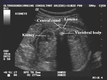
Ultrasound field of a plane and a concave transducer (left) and of multiarray transducers, electronically focused for short and far distances coronal scan of the left upper quadrant shows a cystic lesion (c) of approximately 2 cm in the pancreatic tail.
•prescribe plane perpendicular to coronal plane (©). Place the probe between iliac crest and the lower costal margin to examine in the coronal plane. This case demonstrates the utility of 3d ultrasound, using the coronal plane as a reference point, for patients with an iud who have pelvic pain. Note lobulating outer margin of the lesion (s, spleen;